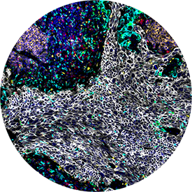
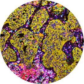
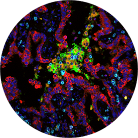
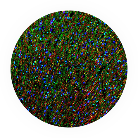
Ultrahigh-plex Proteomics Reveal Brain Cell
Phenotypes and Disease Pathology in Spatial Context
Oligo2
Iba1
MAP2
Synaptophysin
Neurofilament-L
GFAP
MBP
Normal
Alzheimer’s disease
Glioblastoma
Normal
Alzheimer’s disease
Glioblastoma
Normal
Alzheimer’s disease
Glioblastoma
Normal
Alzheimer’s disease
Glioblastoma
Normal
Alzheimer’s disease
Glioblastoma
Normal
Alzheimer’s disease
Glioblastoma
Normal
Alzheimer’s disease
Glioblastoma
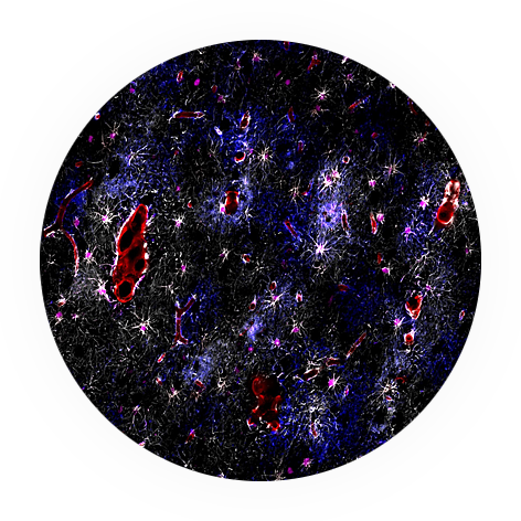
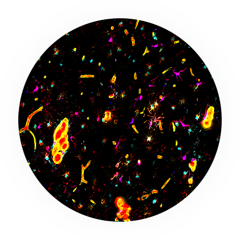
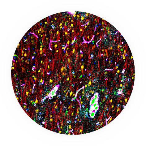
Multiplex images of normal, Alzheimer’s disease (AD), and glioblastoma (GBM) samples showing the expression of five markers.
Lorem Ipsum Dolor Sit Amet
Whether investigating neurodegenerative disease, neuroinflammation, or neuroplasticity, the PhenoCode Discovery Human Neuro Panel empowers you to understand key molecular and cellular interactions in the brain spatially – insights that traditional methods can’t deliver.
Cellular Characterization
Identify and distinguish neuronal subtypes, glial cells, and vascular components with a well-defined set of 42 biomarkers.
Disease-specific Insights
Detect neurodegenerative protein aggregates associated with Alzheimer’s disease and Parkinson’s disease, track inflammation-driven pathology, and assess blood-brain barrier integrity disruptions.
High-impact Data
Use an optimized, ready-to-use panel to spatially phenotype human brain formalin-fixed paraffin-embedded tissues.
Visualize the Complex Cells, Structures, and Interactions of the Brain
Neurons
Neurofilament
MAP2
NeuN
SMA
DAPI
Astroglia
GFAP (GFAP in white)
AQP4
Vimentin
S100B
Microglia
GFAP
IBA1
Vimentin
CD74
Neurons and Glia
GFAP
IBA1
MAP2
NeuN
Vimentin
S100B
Case Studies Highlighting Spatial Proteomics to Study the Brain
Understanding the Brain: Advancing Neurodegeneration Research with Ultrahigh-Plex Spatial Proteomics
Ritu Mihani, PhD
Director of Marketing, Akoya Biosciences
Applying spatial biology to neuroscience research presents unique challenges due to limited tissue availability and the fragile nature of brain tissue.
Learn how ultrahigh-plex spatial phenotyping has been applied to neurodegenerative disease research.
Decoding Neurodegeneration: High-plex Spatial Proteomics Reveal Distinct Cell Phenotypes and Pathological Signatures in Parkinson’s and Alzheimer’s Diseases
Dr. Birgitt Schuele
Associate Professor (Research) of Pathology at Stanford University
Dr. Birgitt Schuele showcases how ultrahigh-plex spatial proteomics is being used to uncover distinct cell phenotypes and pathological signatures in brain tissue.
Learn how spatial biology is advancing our understanding of neurodegeneration and enabling new insights into disease progression.